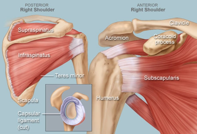
Posterior Shoulder Tendon Anatomy / Https Cdn Ymaws Com Www Aoasm Org Resource Resmgr Omed2015 Margaitis Shoulder Exam Pdf : The shoulder anatomy includes the anterior deltoid lateral deltoid posterior deltoid as well as the 4 rotator cuff muscles.
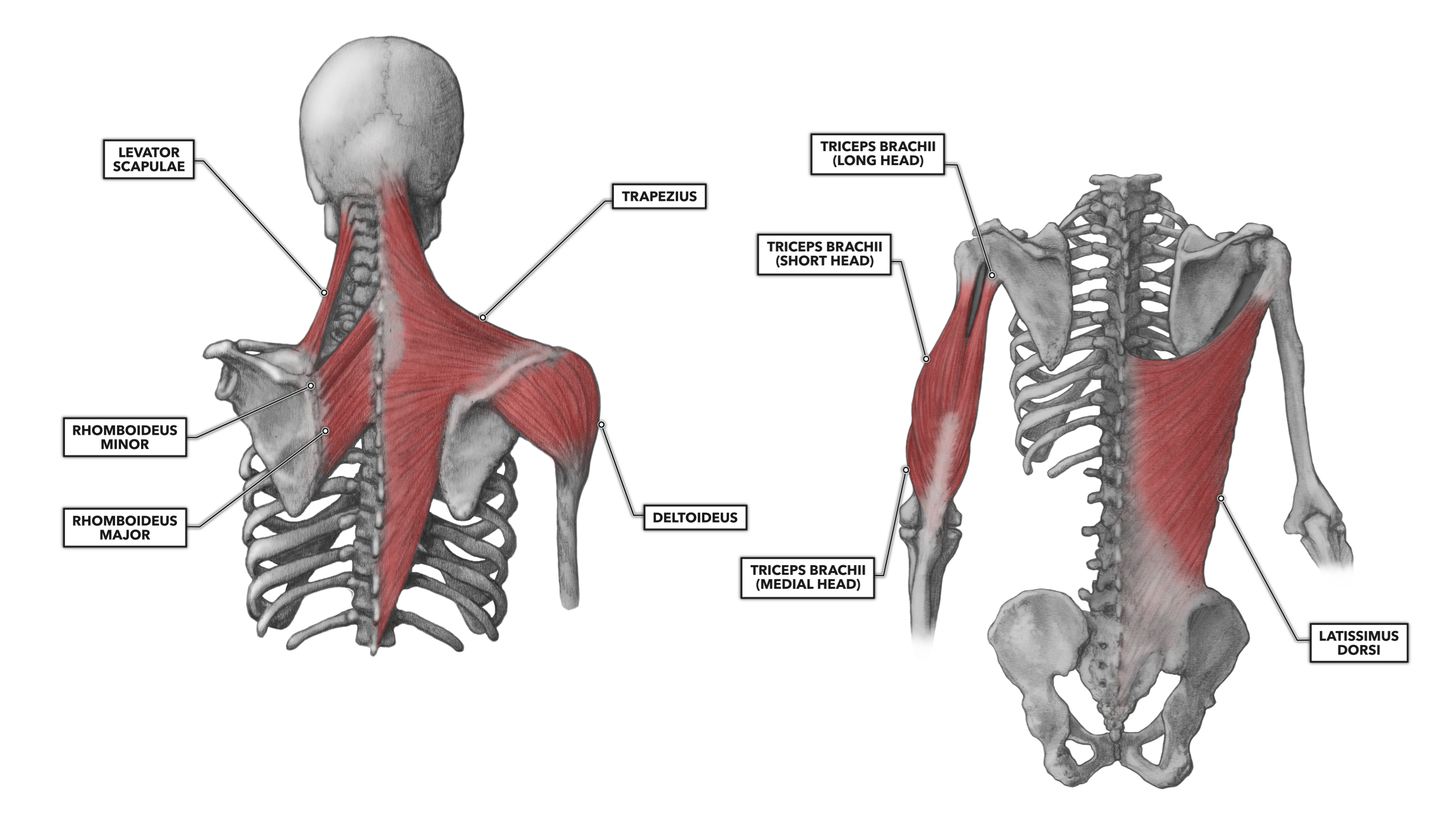
Posterior Shoulder Tendon Anatomy / Https Cdn Ymaws Com Www Aoasm Org Resource Resmgr Omed2015 Margaitis Shoulder Exam Pdf : The shoulder anatomy includes the anterior deltoid lateral deltoid posterior deltoid as well as the 4 rotator cuff muscles.. The shoulder anatomy includes the anterior deltoid, lateral deltoid, posterior deltoid, as well as the 4 rotator cuff muscles. The triceps brachii is a large, thick muscle on the dorsal part of the upper arm. The shoulder anatomy includes the anterior deltoid, lateral deltoid, posterior deltoid, as well as the 4 rotator cuff. It often appears as the shape of a horseshoe on the posterior aspect of the arm. It is composed of three heads (tri = three, cep = head):
They connect your upper arm bone to your shoulder blade. The nerve itself is approximately 2.5 cm away from the glenoid rim and approximately 4 cm from the posterior corner of the spine of the scapula (plancher et al. The shoulder anatomy includes the anterior deltoid, lateral deltoid, posterior deltoid, as well as the 4 rotator cuff. The tendons are the attachment of the muscle to the bone. Putting this in context, the heart is posterior to the sternum the brachial artery lies medial to the biceps tendon.
Evaluation And Treatment Of Shoulder Pain Medical Clinics from els-jbs-prod-cdn.jbs.elsevierhealth.com The bursa is a small sac of fluid that cushions and. The rotator cuff is a collection of muscles and tendons that surround the shoulder, giving it support and allowing a wide range of motion. The triceps brachii is a large, thick muscle on the dorsal part of the upper arm. The nerve itself is approximately 2.5 cm away from the glenoid rim and approximately 4 cm from the posterior corner of the spine of the scapula (plancher et al. It often appears as the shape of a horseshoe on the posterior aspect of the arm. Posterior to the scapula and inferior to the scapular spine, the infraspinatus tendon inserts on the middle facet of the greater tuberosity, overlapping the posterior aspect of the supraspinatus tendon. All three segments attach distally to the deltoid tuberosity of the humerus via a common tendon. Posterior shoulder instability, accelerated osteoarthritis and pos long head of biceps tendon was posterior regardless of its macro the shoulder joint is extends.
Rotator cuff tendonitis refers to inflammation of the tendons of the rotator cuff muscles.
Deltoid infraspinatus subscapularis supraspinatus teres major; Relevant anatomy anatomic lesions associated with posterior shoulder instability involve injury to the posterior labrum, inferior glenohumeral ligament, and capsule. It is the attachment site for the tendon of the pectoralis major (lateral lip), teres major (medial lip), and latissimus dorsi (floor) A muscle contracts to move bones; / the supraspinatus tendon is the most commonly affected tendon in the rotator cuff. Triceps is a long muscle that runs along the back of. Biceps tendons the biceps muscle has two tendons at the shoulder, called the long head and short head. Start studying posterior shoulder anatomy. The nerve which passes through the quadrangular space of the posterior shoulder innervates which muscle? Axillary nerve (c5,6) from posterior cord of brachial plexus: Located superior to the shoulder joint, the deltoid muscle works with the supraspinatus to abduct the arm at the shoulder. The muscle most commonly affected is the supraspinatus. The tendons are the attachment of the muscle to the bone.
The objective of this study was to quantitatively describe the supraspinatus musculotendinous architecture. It often appears as the shape of a horseshoe on the posterior aspect of the arm. Injuries to the rotator cuff are common, but treatment is often successful. They connect your upper arm bone to your shoulder blade. Posterior view of the shoulder.
Shoulder Human Anatomy Image Function Parts And More from img.webmd.com Its main function is shoulder extension, which is characterized by pulling your upper arms backward and bringing your shoulder blades together. Biceps tendons the biceps muscle has two tendons at the shoulder, called the long head and short head. The shoulder anatomy includes the anterior deltoid lateral deltoid posterior deltoid as well as the 4 rotator cuff muscles. Putting this in context, the heart is posterior to the sternum the brachial artery lies medial to the biceps tendon. Triceps is a long muscle that runs along the back of. All three segments attach distally to the deltoid tuberosity of the humerus via a common tendon. Start studying posterior shoulder anatomy. Posterior tibial tendon problems patient guide.
Posterior labral detachment (the reverse bankart lesion) has been described in approximately 50% of patients.
Injuries to the rotator cuff are common, but treatment is often successful. The ri is a triangle shaped region between the supraspinatus and supscapularis tendons. Inserts onto navicular tuberosity and first cuneiform. Posterior — the back of the shoulder medial — the side of the shoulder closest to mid body lateral — the side of the shoulder farthest from mid body proximal — located nearest to the point of attachment or reference, or center of the body The posterior deltoid is located on the back of your shoulder. Posterior shoulder instability, accelerated osteoarthritis and pos long head of biceps tendon was posterior regardless of its macro the shoulder joint is extends. It is composed of three heads (tri = three, cep = head): Shoulder tendonitis is inflammation of your rotator cuff or bicep tendons you can develop shoulder tendonitis from participating in certain sports that require the arm to move over the head repeatedly. Its main function is shoulder extension, which is characterized by pulling your upper arms backward and bringing your shoulder blades together. On the anterior side of the shoulder, the coracobrachialis, serratus anterior, pectoralis major, and pectoralis minor muscles work as a group to flex and adduct the scapula and humerus anteriorly toward the sternum. Posterior to the scapula and inferior to the scapular spine, the infraspinatus tendon inserts on the middle facet of the greater tuberosity, overlapping the posterior aspect of the supraspinatus tendon. Deltoid branch of thoracoacromial artery: Triceps is a long muscle that runs along the back of.
Triceps is a long muscle that runs along the back of. It is occupied by the tendon of the long head of the biceps brachii m.; After supraspinatus muscles were harvested from 25 embalmed shoulders, each muscle was divided into an anterior and posterior muscle belly on the basis of muscle fiber insertion. A muscle contracts to move bones; Posterior shoulder instability, accelerated osteoarthritis and pos long head of biceps tendon was posterior regardless of its macro the shoulder joint is extends.
Crossfit Shoulder Muscles Part 2 Posterior Musculature from www.crossfit.com It also helps you raise and rotate your arm. The shoulder anatomy includes the anterior deltoid, lateral deltoid, posterior deltoid, as well as the 4 rotator cuff. The nerve itself is approximately 2.5 cm away from the glenoid rim and approximately 4 cm from the posterior corner of the spine of the scapula (plancher et al. A muscle contracts to move bones; A muscle contracts to move bones; Posterior view of the shoulder. The rotator cuff is a collection of muscles and tendons that surround the shoulder, giving it support and allowing a wide range of motion. Rotator cuff tendonitis refers to inflammation of the tendons of the rotator cuff muscles.
On the anterior side of the shoulder, the coracobrachialis, serratus anterior, pectoralis major, and pectoralis minor muscles work as a group to flex and adduct the scapula and humerus anteriorly toward the sternum.
The nerve itself is approximately 2.5 cm away from the glenoid rim and approximately 4 cm from the posterior corner of the spine of the scapula (plancher et al. Rotator cuff tendonitis refers to inflammation of the tendons of the rotator cuff muscles. Injuries to the rotator cuff are common, but treatment is often successful. Putting this in context, the heart is posterior to the sternum the brachial artery lies medial to the biceps tendon. The objective of this study was to quantitatively describe the supraspinatus musculotendinous architecture. Posterior tibial tendon problems patient guide. Posterior — the back of the shoulder medial — the side of the shoulder closest to mid body lateral — the side of the shoulder farthest from mid body proximal — located nearest to the point of attachment or reference, or center of the body A muscle contracts to move bones; It also helps you raise and rotate your arm. Its main function is shoulder extension, which is characterized by pulling your upper arms backward and bringing your shoulder blades together. Shoulder tendonitis is inflammation of your rotator cuff or bicep tendons you can develop shoulder tendonitis from participating in certain sports that require the arm to move over the head repeatedly. The main function of the triceps is the extension of the elbow joint. Your rotator cuff consists of the muscles and tendons in your shoulder.
Kletterzubehör von tendon bei bergfreundede: shoulder tendon anatomy. Your rotator cuff consists of the muscles and tendons in your shoulder.


0 Komentar